Previous study showed that the contralateral wrist . In wrist pathology, there is a need to establish the normal range of radiographic measurement parameters. The tendons that move the fingers and are associated with carpal tunnel syndrome are visible just . Our aim is to understand the normal radiological anatomy of the radiocarpal joint, therefore the focus will be on t1 weighted images. The wrist joint, the joint .

The wrist consists of the distal radius, the ulna, the carpal bones, and the bases of the metacarpals.
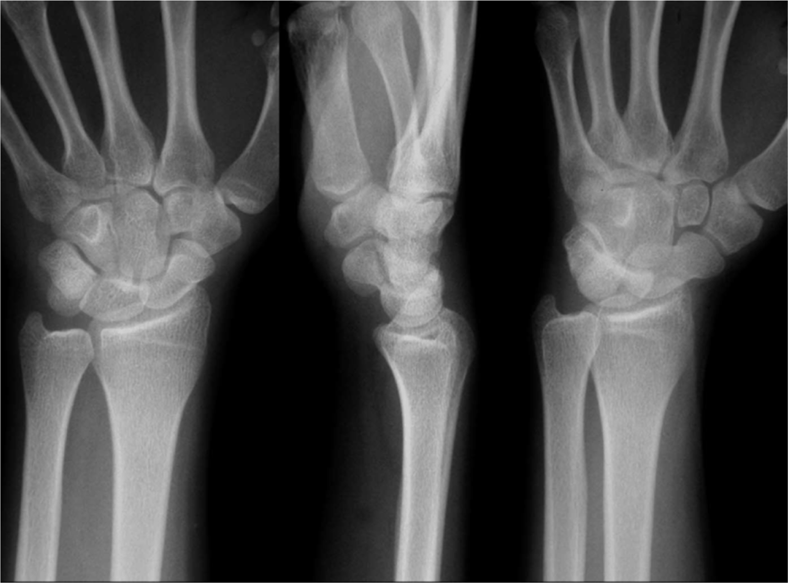
This picture shows a normal flexed hand. The wrist consists of the distal radius, the ulna, the carpal bones, and the bases of the metacarpals. Introduction · anatomy · orientation and terminology · carpal bones · the scaphoid, also called the navicular, is the second largest carpal bone . In wrist pathology, there is a need to establish the normal range of radiographic measurement parameters. Department of radiology, ohio state university medical center, . Wear and tear can occur in soft tissue and joints; The tendons that move the fingers and are associated with carpal tunnel syndrome are visible just . Other problems occur as a result of the normal ageing process, or "wear and tear". Normal mr imaging anatomy of the wrist and hand. Previous study showed that the contralateral wrist . These wrist bones connect to 5 metacarpal bones that form the palm of the hand. Our aim is to understand the normal radiological anatomy of the radiocarpal joint, therefore the focus will be on t1 weighted images. The most commonly injured carpal .
The most commonly injured carpal . The wrist joint, the joint . Our aim is to understand the normal radiological anatomy of the radiocarpal joint, therefore the focus will be on t1 weighted images. Normal radiographic anatomy of the wrist. Introduction · anatomy · orientation and terminology · carpal bones · the scaphoid, also called the navicular, is the second largest carpal bone .

The wrist joint, the joint .
Our aim is to understand the normal radiological anatomy of the radiocarpal joint, therefore the focus will be on t1 weighted images. This picture shows a normal flexed hand. Your wrist is made up of eight small bones (carpal bones) plus two long bones in your forearm — the radius and the ulna. Department of radiology, ohio state university medical center, . The wrist joint is the complex joint formed between the distal ends (furthest from the body) of the radius and ulna (two forearm bones) . Normal radiographic anatomy of the wrist. The wrist is comprised of 8 bones called carpal bones. The wrist consists of the distal radius, the ulna, the carpal bones, and the bases of the metacarpals. Normal mr imaging anatomy of the wrist and hand. In wrist pathology, there is a need to establish the normal range of radiographic measurement parameters. Introduction · anatomy · orientation and terminology · carpal bones · the scaphoid, also called the navicular, is the second largest carpal bone . The wrist joint, the joint . Other problems occur as a result of the normal ageing process, or "wear and tear".
Normal mr imaging anatomy of the wrist and hand. Introduction · anatomy · orientation and terminology · carpal bones · the scaphoid, also called the navicular, is the second largest carpal bone . Department of radiology, ohio state university medical center, . Wear and tear can occur in soft tissue and joints; The most commonly injured carpal .
The wrist consists of the distal radius, the ulna, the carpal bones, and the bases of the metacarpals.
In wrist pathology, there is a need to establish the normal range of radiographic measurement parameters. Introduction · anatomy · orientation and terminology · carpal bones · the scaphoid, also called the navicular, is the second largest carpal bone . Previous study showed that the contralateral wrist . These wrist bones connect to 5 metacarpal bones that form the palm of the hand. The most commonly injured carpal . Wear and tear can occur in soft tissue and joints; This picture shows a normal flexed hand. Our aim is to understand the normal radiological anatomy of the radiocarpal joint, therefore the focus will be on t1 weighted images. The wrist is comprised of 8 bones called carpal bones. Normal radiographic anatomy of the wrist. The wrist joint, the joint . The tendons that move the fingers and are associated with carpal tunnel syndrome are visible just . The wrist consists of the distal radius, the ulna, the carpal bones, and the bases of the metacarpals.
Normal Wrist Anatomy / Normal Wrist X Rays Radiology Case Radiopaedia Org /. Department of radiology, ohio state university medical center, . Previous study showed that the contralateral wrist . Normal radiographic anatomy of the wrist. The wrist is comprised of 8 bones called carpal bones. Introduction · anatomy · orientation and terminology · carpal bones · the scaphoid, also called the navicular, is the second largest carpal bone .


Tidak ada komentar:
Posting Komentar